Distinct transcriptomic biomarkers and pathways associated with cardiovascular and neurovascular dysregulation in long COVID-19 brain fog
Keywords:
Long COVID-19, SARS-CoV-2, Transcriptomics, PathwaysAbstract
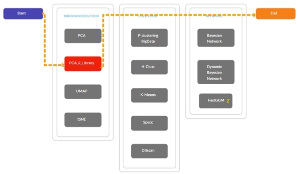
Background: Long COVID-19 often manifests as persistent cognitive impairments, commonly referred to as brain fog, with poorly understood biological mechanisms. Previous studies highlighted neuroinflammation and blood-brain barrier disruption. Here, we identify distinct biomarkers and dysregulated pathways, with a focus on cardiovascular and neurovascular involvement. Methods: We analyzed public RNA-sequencing data (GEO: GSE251849) comparing long COVID-19 patients with brain fog, long COVID-19 patients without cognitive symptoms, convalescent patients, and healthy controls. Differential gene expression, principal component analysis, and pathway enrichment analyses were performed using DESeq2, KEGG, GO, and Enrichr tools. Results: Our analysis revealed a distinct gene expression profile in patients with brain fog, identifying exclusive biomarkers such as NRCAM, GRIN2C, NOS2, and EDNRB. Pathway analysis revealed significant dysregulation in cardiovascular, calcium signaling, and relaxin signaling pathways, suggesting novel biological mechanisms that contribute to cognitive impairment. These findings expand beyond neuroinflammation and highlight potential cardiovascular involvement in long COVID-related cognitive dysfunction. Conclusion: Our study reveals novel transcriptomic signatures that highlight cardiovascular and neurovascular dysregulation, providing promising diagnostic biomarkers and therapeutic targets. The present analysis is exploratory and based on a small cohort (n = 23, subgroups n = 5–7), so findings should be considered preliminary and require validation in larger, independent cohorts.
References
1. Rodriguez-Morales AJ, Bonilla-Aldana DK, Tiwari R, Sah R, Rabaan AA, Dhama K. COVID-19, an Emerging coronavirus infection: Current scenario and recent developments – An overview. J Pure Appl Microbiol. 2020; 14: 05–12.
2. Kishor RS, Ramhari BM. Introduction to Covid-19. Res J Sci Technol. 2020; 12: 338–345.
3. Hon KL, Leung KKY, Leung AK, Qian SY, Chan VP, Ip P, et al. Coronavirus disease 2019 (COVID-19): latest developments in potential treatments. Drugs Context. 2020; 9: 1–14.
4. Rao V, Bhattamisra SK. Recent developments and opportunities in fighting COVID-19. Coronaviruses. 2020; 2.
5. Vishal K, Waghmare SU, Kodage PB. COVID-19 A comprehensive review of signs, symptoms, diagnosis, and treatment strategies. Int J Adv Res Sci Commun Technol. 2024: 51–68.
6. Golzardi M, Hromić-Jahjefendić A, Šutković J, Aydin O, Ünal-Aydın P, Bećirević T, et al. The Aftermath of COVID-19: Exploring the Long-Term Effects on Organ Systems. Biomedicines. 2024; 12: 913.
7. Bellanti JA. The new frontier: Clinical consequences of long COVID. J Precision Respirat Med. 2023; 6: 2–9.
8. Desai AD, Lavelle M, Boursiquot BC, Wan EY. Long-term complications of COVID-19. AJP Cell Physiol. 2021; 322: C1–C11.
9. Lee CCE, Ali K, Connell D, Mordi IR, George J, Lang EM, et al. COVID-19-Associated cardiovascular complications. Diseases. 2021; 9: 47.
10. Van Der Feltz-Cornelis C, Turk F, Sweetman J, Khunti K, Gabbay M, Shepherd J, et al. Prevalence of mental health conditions and brain fog in people with long COVID: A systematic review and meta-analysis. General Hosp Psych. 2024; 88: 10–22.
11. Mathew B, Kumar R, Harilal S, Pappachan LK. Current Perspective of COVID-19 on Neurology: A Mechanistic Insight. Combinat Chem High Throughput Screen. 2021; 25: 763–767.
12. Möller M, Borg K, Janson C, Lerm M, Normark J, Niward K. Cognitive dysfunction in post‐COVID‐19 condition: Mechanisms, management, and rehabilitation. J Internal Med. 2023; 294: 563–581.
13. Krishnan K, Lin Y, Prewitt K-RM, Potter DA. Multidisciplinary Approach to Brain fog and related Persisting Symptoms Post COVID-19. J Health Service Psychol. 2022; 48: 31–38.
14. Su S, Zhao Y, Zeng N, Liu X, Zheng Y, Sun J, et al. Epidemiology, clinical presentation, pathophysiology, and management of long COVID: an update. Mol Psychiatry. 2023; 28: 4056–4069.
15. Grover S, Sahoo S, Mishra E, Gill KS, Mehra A, Nehra R, et al. Fatigue, perceived stigma, self-reported cognitive deficits and psychological morbidity in patients recovered from COVID-19 infection. Asian J Psychiatry. 2021; 64: 102815.
16. Ceban F, Ling S, Lui LMW, Lee Y, Gill H, Teopiz KM, et al. Fatigue and cognitive impairment in Post-COVID-19 Syndrome: A systematic review and meta-analysis. Brain Behav Immun. 2021; 101: 93–135.
17. Talkington GM, Kolluru P, Gressett TE, Ismael S, Meenakshi U, Acquarone M, et al. Neurological sequelae of long COVID: a comprehensive review of diagnostic imaging, underlying mechanisms, and potential therapeutics. Front Neurol. 2025; 15.
18. Greene C, Connolly R, Brennan D, Laffan A, O’Keeffe E, Zaporojan L, et al. Blood–brain barrier disruption and sustained systemic inflammation in individuals with long COVID-associated cognitive impairment. Nat Neurosci. 2024; 27: 421–432.
19. Fernández-Castañeda A, Lu P, Geraghty AC, Song E, Lee M-H, Wood J, et al. Mild respiratory COVID can cause multi-lineage neural cell and myelin dysregulation. Cell .2022; 185: 2452-2468.e16.
20. Missailidis D, Ebrahimie E, Dehcheshmeh MM, Allan C, Sanislav O, Fisher P, et al. A blood-based mRNA signature distinguishes people with Long COVID from recovered individuals. Front Immunol. 2024; 15. doi:10.3389/fimmu.2024.1450853.
21. Kayalar O, Cetinkaya PD, Eldem V, Baris SA, Kokturk N, Kuralay SC, et al. Comparative Transcriptomic Analyses of Peripheral Blood Mononuclear Cells of COVID-19 Patients without Pneumonia and with Severe Pneumonia in the First Year of Follow-Up. Viruses. 2024; 16: 1211.
22. Ye Y, Chen Z, Jiang S, Jia F, Li T, Lu X, et al. Single-cell profiling reveals distinct adaptive immune hallmarks in MDA5+ dermatomyositis with therapeutic implications. Nat Commun. 2022; 13.
23. Ma J, Bai H, Gong T, Mao W, Nie Y, Zhang X et al. Novel skewed usage of B-cell receptors in COVID-19 patients with various clinical presentations. Immunology Letters 2022; 249: 23–32.
24. Lee J-A, Gao F-B. Neuronal functions of ESCRTs. Exp Neurobiol. 2012; 21: 9–15.
25. Ju Y, Bai H, Ren L, Zhang L. The role of Exosome and the ESCRT pathway on enveloped virus infection. International J Mol Sci. 2021; 22: 9060.
26. Ohno M, Sasaki M, Orba Y, Sekiya T, Masum MdA, Ichii O, et al. Abnormal Blood Coagulation and Kidney Damage in Aged Hamsters Infected with Severe Acute Respiratory Syndrome Coronavirus 2. Viruses. 2021; 13: 2137.
27. Tugarov Y, Huet A, Dvorshchenko K. Expression of Lrp1 and Olr1 genes in the blood of patientswith osteoarthritis after Sars-CoV-2 infection. Bull Taras Shevchenko Nat Univ Kyiv Series Biol. 2023; 94: 35–40.
28. Serpente M, Fenoglio C, Villa C, Cortini F, Cantoni C, Ridolfi E, et al. Role of OLR1 and its regulating HSA-MIR369-3P in Alzheimer’s Disease: Genetics and Expression analysis. J Alzheimer Dis. 2011; 26: 787–793.
29. Saini G, Aneja R. Cancer as a prospective sequela of long COVID‐19. BioEssays. 2021; 43.
30. Kavanagh E. Long Covid brain fog: a neuroinflammation phenomenon? Oxford Open Immunol. 2022; 3.
31. Majeed A, Shajar MA. Is hemoglobin the missing link in the pathogenesis of COVID-19? Anaesthesia Pain Intensive Care. 2020; 24: 9–12.
32. Dai L, Jin X, Liu Z. Prognostic and clinicopathological significance of GPRC5A in various cancers: A systematic review and meta-analysis. PLoS ONE. 2021; 16: e0249040.
33. González P a. I, Valdivieso ÁG, Santa-Coloma TA. The G protein-coupled receptor GPRC5A-a phorbol ester and retinoic acid-induced orphan receptor with roles in cancer, inflammation, and immunity. Biochem Cell Biol. 2023; 101: 465–480.
34. Jiang X, Xu X, Wu M, Guan Z, Su X, Chen S, et al. GPRC5A: an emerging biomarker in human cancer. BioMed Res Int. 2018; 2018: 1–11.
35. Cheng Z, Zhang C, Zhao F, Piao J, Cui R, Li B. RASD2 mediates acute Fasting-Induced Antidepressant-Like effects via dopamine D2 receptor activation in ovariectomized mice. Int J Neuropsychopharmacol. 2022; 26: 217–229.
36. Vitucci D, Di Giorgio A, Napolitano F, Pelosi B, Blasi G, Errico F, et al. RASD2 modulates Prefronto-Striatal phenotypes in humans and ‘Schizophrenia-Like behaviors’ in mice. Neuropsychopharmacol. 2015; 41: 916–927.
37. Patel MA, Knauer MJ, Nicholson M, Daley M, Van Nynatten LR, Martin C, et al. Elevated vascular transformation blood biomarkers in Long-COVID indicate angiogenesis as a key pathophysiological mechanism. Mol Med. 2022; 28.
38. Lieberman N a. P, Peddu V, Xie H, Shrestha L, Huang M-L, Mears MC, et al. In vivo antiviral host transcriptional response to SARS-CoV-2 by viral load, sex, and age. PLoS Biol. 2020; 18: e3000849.
39. Shaghaghi MA, Murphy B, Eck P. The SLC2A14 gene: genomic locus, tissue expression, splice variants, and subcellular localization of the protein. Biochem Cell Biol. 2016; 94: 331–335.
40. Henderson-Smith A, Chow D, Meechoovet B, Aziz M, Jacobson SA, Shill HA, et al. SMG1 Identified as a Regulator of Parkinson’s Disease-Associated alpha-Synuclein through siRNA Screening. PLoS ONE. 2013; 8: e77711.
41. Chatys-Bogacka Ż, Mazurkiewicz I, Słowik J, Słowik A, Drabik L, Wnuk M. Association between brain fog, cardiac injury, and quality of life at work after hospitalization due to COVID-19. Medycyna Pracy. 2024; 75(1): 3-17.
42. Mailian DE, Kolomiets VV. The role of calcium metabolism dysregulation in the pathogenesis of cardiovascular diseases. Russ J Cardiol. 2019: 78–85.
43. Schrank S, Barrington N, Stutzmann GE. Calcium-Handling defects and neurodegenerative disease. Cold Spring Harbor Perspect Biol. 2019; 12: a035212.
44. Fontes-Dantas FL, Fernandes GG, Gutman EG, De Lima EV, Antonio LS, Hammerle MB, et al. SARS-CoV-2 Spike protein induces TLR4-mediated long-term cognitive dysfunction recapitulating post-COVID-19 syndrome in mice. Cell Rep. 2023; 42: 112189.
45. Low RN, Low RJ, Akrami A. A review of cytokine-based pathophysiology of Long COVID symptoms. Front Med. 2023; 10.
46. Zadeh AF, Arnsten AFT, Wang M. Scientific Rationale for the Treatment of Cognitive Deficits from Long COVID. Neurol Int. 2023; 15: 725–742.
47. Gupta SC, Ravikrishnan A, Liu J, Mao Z, Pavuluri R, Hillman BG, et al. The NMDA receptor GluN2C subunit controls cortical excitatory-inhibitory balance, neuronal oscillations and cognitive function. Scientific Reports 2016; 6. doi:10.1038/srep38321.
48. Rubino E, Italia M, Giorgio E, Boschi S, Dimartino P, Pippucci T, et al. Exome sequencing reveals a rare damaging variant in GRIN2C in familial late-onset Alzheimer’s disease. Alzheimer Res Ther. 2025; 17.
49. Alcendor DJ. Dysregulation of endothelin-1: Implications for health disparities in Alzheimer’s disease. J Personal Med. 2020; 10: 199.
50. Koyama Y. Endothelin ETB Receptor-Mediated Astrocytic Activation: Pathological roles in brain Disorders. Int J Mol Sci. 2021; 22: 4333.
51. Michinaga S, Hishinuma S, Koyama Y. Roles of astrocytic endothelin ETB receptor in traumatic brain injury. Cells. 2023; 12: 719.
52. Cespuglio R, Strekalova T, Spencer PS, Román GC, Reis J, Bouteille B, et al. SARS-CoV-2 infection and sleep disturbances: nitric oxide involvement and therapeutic opportunity. Sleep. 2021; 44.
53. Chen BP, Lane TE. Lack of nitric oxide synthase type 2 (NOS2) results in reduced neuronal apoptosis and mortality following mouse hepatitis virus infection of the central nervous system. J NeuroVirol. 2002; 8: 58–63.
54. Garvin RA. Relaxin levels in matched sera and CSF from subjects with multiple sclerosis. Ital J Anat Embryol. 2018; 123: 130–135.
55. Chow BSM, Chew EGY, Zhao C, Bathgate RD, Hewitson TD, Samuel CS. Relaxin Signals through a RXFP1-pERK-nNOS-NO-cGMP-Dependent Pathway to Up-Regulate Matrix Metalloproteinases: The Additional Involvement of iNOS. PLoS ONE. 2012; 7: e42714.
56. Baccari M, Bani D. Relaxin and nitric oxide signalling. Curr Protein Peptide Sci. 2008; 9: 638–645.
57. Hoos MD, Vitek MP, Ridnour LA, Wilson J, Jansen M, Everhart A, et al. The impact of human and mouse differences in NOS2 gene expression on the brain’s redox and immune environment. Mol Neurodegener. 2014; 9.
58. Riquelme P, Amodio G, Macedo C, Moreau A, Obermajer N, Brochhausen C, et al. DHRS9 is a stable marker of human regulatory macrophages. Transplantation. 2017; 101: 2731–2738.
59. Moy SS, Nonneman RJ, Young NB, Demyanenko GP, Maness PF. Impaired sociability and cognitive function in Nrcam-null mice. Behav Brain Res. 2009; 205: 123–131.
60. Reiken S, Sittenfeld L, Dridi H, Liu Y, Liu X, Marks AR. Alzheimer’s‐like signaling in brains of COVID‐19 patients. Alzheimer Dementia. 2022; 18: 955–965.
61. Iadecola C, Yaffe K, Biller J, Bratzke LC, Faraci FM, Gorelick PB, et al. Impact of hypertension on cognitive function: a scientific statement from the American Heart Association. Hypertension. 2016; 68.
62. Wolters FJ, Zonneveld HI, Hofman A, Van Der Lugt A, Koudstaal PJ, Vernooij MW, et al. Cerebral perfusion and the risk of dementia. Circulation. 2017; 136: 719–728.
63. Alosco ML, Brickman AM, Spitznagel MB, Garcia SL, Narkhede A, Griffith EY, et al. Cerebral Perfusion is Associated With White Matter Hyperintensities in Older Adults With Heart Failure. Congestive Heart Failure. 2013; 19.
64. Haloot J, Bhavaraju-Sanka R, Pillarisetti J, Verduzco-Gutierrez M. Autonomic dysfunction related to postacute SARS-COV-2 syndrome. Phys Med Rehabil Clin North Am. 2023; 34: 563–572.
65. Pulliam L, Sun B, Mustapic M, Chawla S, Kapogiannis D. Plasma neuronal exosomes serve as biomarkers of cognitive impairment in HIV infection and Alzheimer’s disease. J NeuroVirol. 2019; 25: 702–709.
66. Sun B, Fernandes N, Pulliam L. Profile of neuronal exosomes in HIV cognitive impairment exposes sex differences. AIDS. 2019; 33: 1683–1692.
67. Reinhold D, Farztdinov V, Yan Y, Meisel C, Sadlowski H, Kühn J, et al. The brain reacting to COVID-19: analysis of the cerebrospinal fluid proteome, RNA and inflammation. J Neuroinflam. 2023; 20.
68. Madeira C, Vargas-Lopes C, Brandão CO, Reis T, Laks J, Panizzutti R, et al. Elevated glutamate and glutamine levels in the cerebrospinal fluid of patients with probable Alzheimer’s disease and depression. Front Psych. 2018; 9.
69. López-Hernández Y, Monárrez-Espino J, López DAG, Zheng J, Borrego JC, Torres-Calzada C, et al. The plasma metabolome of long COVID patients two years after infection. Scient Rep. 2023; 13.
70. Li X, Edén A, Malwade S, Cunningham JL, Bergquist J, Weidenfors JA, et al. Central and peripheral kynurenine pathway metabolites in COVID-19: Implications for neurological and immunological responses. Brain Behav Immun. 2024; 124: 163-176.
71. Barnden L, Thapaliya K, Eaton-Fitch N, Barth M, Marshall-Gradisnik S. Altered brain connectivity in Long Covid during cognitive exertion: a pilot study. Front Neurosci. 2023; 17.
72. Sun Y, Sun J, Chen X, Wang Y, Gao X. EEG signatures of cognitive decline after mild SARS-CoV-2 infection: an age-dependent study. BMC Med. 2024; 22.
73. Crosier R, Kafil TS, Paterson DI. Imaging for cardiovascular complications of COVID-19: Cardiac Manifestations in context. Canad J Cardiol 2023; 39: 779–792.
74. Rêgo LOS, Braga LLA, Vilas-Boas GS, Cardoso MSO, Duraes AR. Cardiovascular and Neurological Complications of COVID-19: A Narrative review. J Clin Med. 2023; 12: 2819.
75. Grätz C, Bui MLU, Thaqi G, Kirchner B, Loewe RP, Pfaffl MW. Obtaining Reliable RT-QPCR Results in Molecular Diagnostics-MIQE Goals and Pitfalls for transcriptional biomarker discovery. Life. 2022; 12: 386.
76. Gonzalez-Ortiz F, Kac PR, Brum WS, Zetterberg H, Blennow K, Karikari TK. Plasma phospho-tau in Alzheimer’s disease: towards diagnostic and therapeutic trial applications. Mol Neurodegen. 2023; 18.
77. Barro C, Chitnis T, Weiner HL. Blood neurofilament light: a critical review of its application to neurologic disease. Ann Clin Transla Neurol. 2020; 7: 2508–2523.
78. Chapman MA, Arif M, Emanuelsson EB, Reitzner SM, Lindholm ME, Mardinoglu A, et al. Skeletal Muscle Transcriptomic Comparison between Long-Term Trained and Untrained Men and Women. Cell Rep. 2020; 31: 107808.
79. Dadsena R, Walders J, Costa AS, Wetz S, Romanzetti S, Lischewski SA, et al. Two‐year impact of COVID‐19: Longitudinal MRI brain changes and neuropsychiatric trajectories. Psychiatry Clin Neurosci. 2025; 79(4): 176-186.
80. McMullen PD, Pendse SN, Black MB, Mansouri K, Haider S, Andersen ME, et al. Addressing systematic inconsistencies between in vitro and in vivo transcriptomic mode of action signatures. Toxicol in Vitro. 2019; 58: 1–12.
81. Cataldo SA, Micciulli A, Margulis L, Cibeyra M, Defeo S, Horovitz SG, et al. Cognitive impact and brain structural changes in long COVID patients: a cross-sectional MRI study two years post infection in a cohort from Argentina. BMC Neurol. 2024; 24.
Downloads
Published
Issue
Section
License
Copyright (c) 2026 Authors

This work is licensed under a Creative Commons Attribution-NonCommercial-NoDerivatives 4.0 International License.