Molecular xenomonitoring of lymphatic filariasis transmission in Okobo, Akwa Ibom State, Nigeria

Authors

  • N. W. Ikpeida Department of Animal and Environmental Biology, Faculty of Biological Sciences, University of Uyo, Uyo, Akwa Ibom State, Nigeria Author
  • K. N. Opara Department of Animal and Environmental Biology, Faculty of Biological Sciences, University of Uyo, Uyo, Akwa Ibom State, Nigeria Author
  • N. I. Udoidung Department of Animal and Environmental Biology, Faculty of Biological Sciences, University of Uyo, Uyo, Akwa Ibom State, Nigeria Author
  • M. A. Adeleke Department of Zoology, Osun State University, Osogbo, Nigeria; Federal University of Health, Ila Oragun, Osun State, Nigeria Author
  • F. M. Chikezie Department of Animal and Environmental Biology, Faculty of Biological Sciences, University of Uyo, Uyo, Akwa Ibom State, Nigeria Author
  • C. A. Yaro Department of Animal and Environmental Biology, Faculty of Biological Sciences, University of Uyo, Uyo, Akwa Ibom State, Nigeria Author
  • Z. O. Iwalewa Department of Zoology, Osun State University, Osogbo, Nigeria Author
  • I. I. Silas Department of Zoology and Environmental Biology, University of Calabar, Cross River State, Nigeria Author

Keywords:

PCR, Lymphatic filariasis, Molecular xenomonitoring, Mosquitoes, Wuchereria bancrofti, Ssp I

Abstract

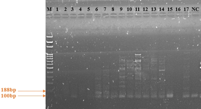
Lymphatic filariasis (LF) is still a significant public health disease of great burden in endemic regions. Molecular xenomonitoring (MX) is crucial to evaluate elimination efforts during mass drug administration (MDA) and post-MDA settings. This study investigated Wuchereria bancrofti prevalence in mosquitoes using molecular techniques in Okobo LGA, an endemic area that has undergone rounds of MDA. Entomological survey involved capturing mosquitoes using CDC light traps during wet (May-July, 2024) and dry (November 2024 - January 2025) seasons. A total of 518 female mosquitoes were captured and morphologically identified comprising of Culex spp. (95.95%, 497) and Anopheles spp. (4.05%, 21). PCR was used to detect W. bancrofti DNA in ≤ 30 mosquito pools. This protocol amplified a segment of the Ssp I repeat found in all larva stages of W. bancrofti enabling detection of parasite’s DNA. Results showed 82.22% pool positivity. PoolTestR was used to estimate prevalence of W. bancrofti in mosquitoes throughout the LGA using Maximum Likelihood Estimates (MLE). The overall estimated prevalence (MLE) of W. bancrofti DNA in Culex spp. mosquitoes was 9.99% (95% CI: 6.49%-14.69%) while Anopheles spp., was 63.91% (95% CI: 39.59%–84.67%). The high infection rate revealed by this study begs the need for continued MDA in Okobo as high estimated prevalence is suggestive of ongoing human infection. These findings provide a baseline data for LF monitoring and elimination strategies and emphasizes the importance of molecular surveillance in transmission assessment surveys in endemic LGAs.

DOI: http://dx.doi.org/10.5281/zenodo.21250014

References

1. WHO. Global Programme to Eliminate Lymphatic Filariasis: Progress Report 2022.Weekly Epidemiological Record Relevé Épidémiologique Hebdomadaire.; 2023. Accessed: November 26, 2023. https://mectizan.org/wp-content/uploads/2023/10/WER9841-eng-fre.pdf

2. Curran DM, Grote A, Nursimulu N, Geber A, Voronin D, Jones DR, et al. Modeling the metabolic interplay between a parasitic worm and its bacterial endosymbiont allows the identification of novel drug targets. eLife. 2020;9:e51850. doi: https://doi.org/10.7554/eLife.51850

3. WHO. Lymphatic Filariasis: Managing Morbidity and Preventing Disability: An Aide-Mémoire for National Programme Managers. World Health Organization; 2021. Accessed: November 26, 2023. https://apps.who.int/iris/handle/10665/339931

4. WHO. Lymphatic filariasis factsheet. World Health Organization (WHO) www.who.int. 2023. Accessed: November 24, 2023. https://www.who.int/news-room/fact-sheets/detail/lymphatic-filariasis#:~:text=WHO%20recommends%20the%20following%20MDA

5. Cross JH. Filarial Nematodes. University of Texas Medical Branch at Galveston; 1996. Accessed: November 24, 2023. https://www.ncbi.nlm.nih.gov/books/NBK7844/

6. Ottesen EA, Hooper PJ, Bradley M, Biswas G. The Global Programme to Eliminate Lymphatic Filariasis: Health Impact after 8 Years. PLoS Negl Trop Dis. 2008;2(10):e317. doi: https://doi.org/10.1371/journal.pntd.0000317

7. Ichimori K, King JD, Engels D, Yajima A, Mikhailov A, Lammie P, Ottesen EA. Global Programme to Eliminate Lymphatic Filariasis: The Processes Underlying Programme Success. PLoS Negl Trop Dis. 2014;8(12):e3328. doi: https://doi.org/10.1371/journal.pntd.0003328

8. WHO. The role of polymerase chain reaction techniques for assessing lymphatic filariasis transmission Report of a workshop cosponsored by the WHO and DBL-Centre for Health Research and Development of Copenhagen. www.who.int. 2009. Accessed: March 5, 2024. https://www.who.int/publications/i/item/WHO-HTM-NTD-PCT-2009.1

9. Eneanya OA, Fronterre C, Anagbogu I, Okoronkwo C, Garske T, Cano J, Donnelly CA. Mapping the baseline prevalence of lymphatic filariasis across Nigeria. Parasites Vectors. 2019;12(1). doi: https://doi.org/10.1186/s13071-019-3682-6

10. Expanded special project for elimination of neglected tropical diseases, ESPEN. Lymphatic Filariasis, Nigeria. 2023. Accessed: November 30, 2023. https://espen.afro.who.int/diseases/lymphatic-filariasis

11. de Souza DK, Koudou B, Kelly-Hope LA, Wilson MD, Bockarie MJ, Boakye DA. Diversity and transmission competence in lymphatic filariasis vectors in West Africa, and the implications for accelerated elimination of Anopheles-transmitted filariasis. Parasites Vectors. 2012;5(1):259. doi: https://doi.org/10.1186/1756-3305-5-259

12. Shekarau E, Uzoanya M, Ogbulafor N; Severe Malaria Working Group. Severe malaria intervention status in Nigeria: workshop meeting report. Malaria J. 2024;23(1):177. doi: https://doi.org/10.1186/s12936024050011

13. Atting IA, Ekpo ND, Akpan ME, Bassey BE, Asuquo MJ, Usip LPE, et al. Insecticide Susceptibility Profile of Malaria Vector Populations from the Coastal and Mainland Areas of Akwa Ibom State, Nigeria. J Adv Med Med Res. 2019;29:1-11. doi: https://doi.org/10.9734/jammr/2019/v29i730099

14. Essien UB, Udoidung NI, Ikpeida NW, Ekpo ND, Ndubuka CE, Ogbonna KD. Susceptibility status of female anopheles’ mosquito to organophosphate and carbamate insecticides in market environs in Itu LGA, Akwa Ibom State, Nigeria. Int J Sci Res Arch. 2024;12(2):2467-2475. doi: https://doi.org/10.30574/ijsra.2024.12.2.1567

15. Pilotte N, Unnasch TR, Williams SA. The Current Status of Molecular Xenomonitoring for Lymphatic Filariasis and Onchocerciasis. Trends Parasitol. 2017;33(10):788-798. doi: https://doi.org/10.1016/j.pt.2017.06.008

16. de Souza DK, Bockarie MJ. Current perspectives in the epidemiology and control of lymphatic filariasis. Clin Microbiol Rev. 2025;38(2):e0012623. doi: https://doi.org/10.1128/cmr.00126-23

17. WHO. Lymphatic Filariasis: A Handbook of Practical Entomology for National Lymphatic Filariasis Elimination Programmes. World Health Organization: Geneva, Switzerland.; 2013. Accessed: November 26, 2023. https://iris.who.int/bitstream/handle/10665/87989/9789241505642_eng.pdf

18. Pedersen EM, Stolk WA, Laney SJ, Michael E. The role of monitoring mosquito infection in the Global Programme to Eliminate Lymphatic Filariasis. Trends Parasitol. 2009;25(7):319-327. doi: https://doi.org/10.1016/j.pt.2009.03.013

19. Rao RU, Atkinson LJ, Ramzy RM, Helmy H, Farid HA, Bockarie MJ, et al. A real-time PCR-based assay for detection of Wuchereria bancrofti DNA in blood and mosquitoes. Am J Trop Med Hyg. 2006;74(5):826-832. doi: https://doi.org/10.4269/ajtmh.2006.74.826

20. Jachowski LA. Filariasis in American Samoa. Am J Epidemiol. 1954;60(2):186-203. doi: https://doi.org/10.1093/oxfordjournals.aje.a119712

21. Suzuki T, Sone F. The bionomics of filariasis vectors in Western Samoa. Eisei Dobutsu. 1974;25(3):251-257. doi: https://doi.org/10.7601/mez.25.251

22. Sherrard-Smith E, Skarp JE, Beale AD, Fornadel C, Norris LC, Moore SJ,, et al. Mosquito feeding behavior and how it influences residual malaria transmission across Africa. Proc Nat Acad Sci USA. 2019;116(30):15086-15095. doi: https://doi.org/10.1073/pnas.1820646116

23. Shannon RC. On the Classification of Brazilian Culicidae with special Reference to those capable of harboring the Yellow Fever Virus. Proc Entomol Soc Washington. 1931;33(6).

24. Edwards FW. Mosquitoes of the Ethiopian Region III. Ann Entomol Soc Am. 1941;35(4):475-475. doi: https://doi.org/10.1093/aesa/35.4.475a

25. Gillet JD. Common African Mosquitoes and Their Medical Importance. William Heinemann Medical Books Ltd.; 1972:106.

26. Prat HD. Keys for Identification of Vectors (Annex 1). World Health Organization (W.H.O); 1972. Accessed: December 16, 2023. http://apps.who.int/iris/bitstream/handle/10665/39005/Vector_Control_in_International_Health_eng.pdf?sequence=1&isAllowed=y

27. QIAGEN®. AllPrep DNA/RNA Mini Handbook; 2020. Accessed: October 6, 2023. https://www.qiagen.com/us/resources/download.aspx?id=580866a6-56c6-4674-8566-2852164d8519&lang=en

28. Zhong M, McCarthy J, Bierwert L, Lizotte-Waniewski M, Chanteau S, Nutman TB, et al. A Polymerase Chain Reaction Assay for Detection of the Parasite Wuchereria bancrofti in Human Blood Samples. Am J Trop Med Hyg. 1996;54(4):357-363. doi: https://doi.org/10.4269/ajtmh.1996.54.357

29. ESRI. ArcGIS. Android application software. 2022. https://play.google.com/store/apps/details?id=com.esri.arcgis

30. ESRI. ArcGIS Desktop. Desktop GIS Software Suite - Esri. Environmental Systems Research Institute (ESRI). 2016. https://www.esri.com/en-us/arcgis/products/arcgis-desktop/overview

31. McLure A, O’Neill B, Mayfield H, Lau C, McPherson B. PoolTestR: An R package for estimating prevalence and regression modelling for molecular xenomonitoring and other applications with pooled samples. Environ Model Software. 2021;145:105158. doi: https://doi.org/10.1016/j.envsoft.2021.105158

32. Lenhart A, Eigege A, Kal A, Pam D, Miri ES, Gerlong G, et al. Contributions of different mosquito species to the transmission of lymphatic filariasis in central Nigeria: implications for monitoring infection by PCR in mosquito pools. Filaria J. 2007;6(1). doi: https://doi.org/10.1186/1475-2883-6-14

33. Okorie PN, McKenzie FE, Ademowo OG, Bockarie M, Kelly-Hope L. Nigeria Anopheles Vector Database: An Overview of 100 Years’ Research. PLoS ONE. 2011;6(12):e28347. doi: https://doi.org/10.1371/journal.pone.0028347

34. Manasseh Msugh-Ter M, Palegh A, Dechi A, Aondowase. Anopheles and Culex Mosquito Species Diversity and Its Epidemiological Implications in the Makurdi Area of Benue State, North Central Nigeria. 2017. https://www.scholarsresearchlibrary.com/articles/anopheles-and-culex-mosquito-species-diversity-and-its-epidemiological-implications-in-the-makurdi-area-of-benue-state-n.pdf

35. WHO. Validation of elimination of lymphatic filariasis as a public health problem. 2017. https://iris.who.int/bitstream/handle/10665/254377/9789241511957-eng.pdf

36. Aliyu AA, Sow GJ, Ndams IS. Entomological Survey of Mosquito Vectors of Lymphatic Filariasis in Talatan-mafara and Tsafe Local Government Areas of Zamfara State, Nigeria. FUDMA J Sci. 2020;4(2):207-216. doi: https://doi.org/10.33003/fjs-2020-0402-213

37. Uttah EC, Wokem GN, Okonofua C. The Abundance and Biting Patterns of Culex quinquefasciatus Say (Culicidae) in the Coastal Region of Nigeria. ISRN Zool. 2013;2013:1-7. doi: https://doi.org/10.1155/2013/640691

38. Odoh VU, Sodang CJ, Olayemi IK, Musa AS, Odoh SS, Tegeri K, Onoja AEA. Distribution, Species Composition and Biting Behaviour of Adult Culex Mosquitoes in Badeggi, Niger State, Nigeria. Dutse J Pure Appl Sci. 2025;11(1b):223-233. doi: https://doi.org/10.4314/dujopas.v11i1b.24

39. Linthicum KJ, Britch SC, Anyamba A. Rift Valley Fever: An Emerging Mosquito-Borne Disease. Annu Rev Entomol. 2016;61(1):395-415. doi: https://doi.org/10.1146/annurev-ento-010715-023819

40. Mohammed BR, Yayo AM, Ajanusi OJ, Lawal IA. Relative abundance and molecular identification of Culex pipiens complex (Diptera: Culicidae), in Kura Local Government Area, North-western Nigeria. Parasite Epidemiol Control. 2021;14:e00213. doi: https://doi.org/10.1016/j.parepi.2021.e00213

41. Adesoye O, Oyeniyi T, Babalola A, Olagundoye O, Izekor R, Adetunji O, et al. Entomological Collections and Identifications of Mosquito Faunas in Selected Area Councils of Nigeria Federal Capital Territory. Lafia J Scient Indust Res. 2024;2(2):134-138. doi: https://doi.org/10.62050/ljsir2024.v2n2.341

42. Burkot T, Ichimori K. The PacELF programme: will mass drug administration be enough? Trends Parasitol. 2002;18(3):109-115. doi: https://doi.org/10.1016/s1471-4922(01)02221-8

43. Rao RU, Samarasekera SD, Nagodavithana KC, Punchihewa MW, Dassanayaka TD, Gamini PKD, et al. Programmatic Use of Molecular Xenomonitoring at the Level of Evaluation Units to Assess Persistence of Lymphatic Filariasis in Sri Lanka. PLOS Negl Trop Dis. 2016;10(5):e0004722-. https://doi.org/10.1371/journal.pntd.0004722

44. Farid HA, Weil GJ, Setouhy ME, Morsy ZS, Ramzy RMR, Helmy H. A Critical Appraisal of Molecular Xenomonitoring as a Tool for Assessing Progress toward Elimination of Lymphatic Filariasis. Am J Trop Med Hyg. 2007;77(4):593-600. doi: https://doi.org/10.4269/ajtmh.2007.77.593

45. Kouassi BL, de Souza DK, Goepogui A, Narh CA, King SA, Mamadou BS, et al. Assessing the presence of Wuchereria bancrofti in vector and human populations from urban communities in Conakry, Guinea. Parasites Vectors. 2015;8(1):492. doi: https://doi.org/10.1186/s130710151077x

46. Rao RU, Samarasekera SD, Nagodavithana KC, Goss CW, Punchihewa MW, Dassanayaka TDM, et al. Comprehensive Assessment of a Hotspot with Persistent Bancroftian Filariasis in Coastal Sri Lanka. Am J Trop Med Hyg. 2018;99(3):735-742. doi: https://doi.org/10.4269/ajtmh.18-0169

47. Schmaedick MA, Koppel AL, Pilotte N, Torres M, Williams SA, Dobson SL, et al. Molecular Xenomonitoring Using Mosquitoes to Map Lymphatic Filariasis after Mass Drug Administration in American Samoa. PLOS Negl Trop Dis. 2014;8(8):e3087-e3087. doi: https://doi.org/10.1371/journal.pntd.0003087

48. Materu GS. Prevalence of Wuchereria bancrofti Infection in Mosquitoes from Pangani District, Northeastern Tanzania | East Africa Science. 2016. Accessed: July 17, 2025. https://easci.eahealth.org/easci/article/view/16/75

49. Bartilol B, Babu L, Garama K, Karisa J, Kamau A, Mwandawiro C, et al. Molecular xenomonitoring reveals Anopheles funestus and An. rivulorum as the primary vectors of lymphatic filariasis in coastal Kenya. Parasites Vectors. 2024;17(1):425. doi: https://doi.org/10.1186/s13071024065130

50. Dorkenoo MA, de Souza DK, Apetogbo Y, Oboussoumi K, Yehadji D, Tchalim M, et al. Molecular xenomonitoring for post-validation surveillance of lymphatic filariasis in Togo: no evidence for active transmission. Parasites Vectors. 2018;11(1). doi: https://doi.org/10.1186/s13071-017-2611-9

51. CDC. Lymphatic Filariasis. 2024. Accessed: February 10, 2025. https://www.cdc.gov/filarial-worms/about/lymphatic-filariasis.html?CDC_AAref_Val=https://www.cdc.gov/parasites/lymphaticfilariasis/index.html

52. Waje T, Iliyasu C, Yaki LM, Auta IK. A review of epidemiology of lymphatic filariasis in Nigeria. Pan African Med J. 2024;47(142). doi: https://doi.org/10.11604/pamj.2024.47.142.39746

53. Erickson SM, Thomsen EK, Keven JB, Vincent N, Koimbu G, Siba PM, et al. Mosquito-Parasite Interactions Can Shape Filariasis Transmission Dynamics and Impact Elimination Programs. PLoS Negl Trop Dis. 2013;7(9):e2433. doi: https://doi.org/10.1371/journal.pntd.0002433

54. Pam DD, de Souza DK, D'Souza S, Opoku M, Sanda S, Nazaradden I, et al. Is mass drug administration against lymphatic filariasis required in urban settings? The experience in Kano, Nigeria. PLOS Negl Trop Dis. 2017;11(10):e0006004. doi: https://doi.org/10.1371/journal.pntd.0006004

55. Luka NE, Bala GW, Dikwa KB, Omalu KB, Ewofere ICJ, Lawal EN, Mamuda BN. Evaluating the Effectiveness of Molecular Xeno Monitoring for Lymphatic Filariasis Surveillance in Low-Prevalence Areas of Kaduna South LGA, Kaduna State, Nigeria. Int J Appl Biol Res. 2024;15(2):162-170. https://journal.futminna.edu.ng/index.php/IJABR/issue/download/128/900

56. Chu BK, Deming M, Biritwum NK, Bougma WR, Dorkenoo AM, El-Setouhy M, et al. Transmission Assessment Surveys (TAS) to Define Endpoints for Lymphatic Filariasis Mass Drug Administration: A Multicenter Evaluation. PLoS Negl Trop Dis. 2013;7(12):e2584. doi: https://doi.org/10.1371/journal.pntd.0002584

57. WHO. Transmission assessment surveys in the global programme to eliminate lymphatic filariasis : WHO position statement. Control of Neglected Tropical Diseases (NTD). 2012. Accessed: July 17, 2025. https://www.who.int/publications/i/item/WHO-HTM-NTD-PCT-2012.9

58. Vasuki V, Krishnamoorthy K, Subramanian S, Sadanandane C, Balasubramaniyan R, Sivagnaname N, et al. Molecular xenomonitoring of Wuchereria bancrofti infection in three different evaluation settings of lymphatic filariasis elimination programme in India. Int J Infect Dis. 2025;152:107807. doi: https://doi.org/10.1016/j.ijid.2025.107807

59. Collyer BS, Irvine MA, Hollingsworth TD, Bradley M, Anderson RM. Defining a prevalence level to describe the elimination of Lymphatic Filariasis (LF) transmission and designing monitoring & evaluating (M&E) programmes post the cessation of mass drug administration (MDA). PLOS Negl Trop Dis. 2020;14(10):e0008644. doi: https://doi.org/10.1371/journal.pntd.0008644

Downloads

Published

2026-03-31

How to Cite

Molecular xenomonitoring of lymphatic filariasis transmission in Okobo, Akwa Ibom State, Nigeria. (2026). European Journal of Biological Research, 16(1), 1-12. https://journals.tmkarpinski.com/index.php/ejbr/article/view/4